
Incubator Accessories




Diagnostic Biosystems CALRETININ H-5 Calretinin (H-5)
Calbindin D28K and Calretinin (also designated CR or 29 kDa Calbindin) are two closely related intracellular calcium-binding proteins belonging to the Troponin-C superfamily. Initially isolated from chick retina. Calretinin is an intracellular calcium-binding protein belonging to the troponin C superfamily characterized by a structural motif described as the EF-hand domain. The immunohistochemical detection of calretinin in developing cerebellum is restricted to the later stages indicated by weak staining from week 21 of gestation in Purkinje and basket cells and in neurons of the dentate nucleus. The intensity of staining increases as the cerebellum matures. In tumors, calretinin has been detected in mesotheliomas and some pulmonary adenocarcinomas.\n


Diagnostic Biosystems IGA-FITC IgA-FITC
This antibody reacts with the alpha chain of human IgA. Traces of contaminating antibodies have been removed by solid phase absorption with human plasma proteins. This antibody is well suited for tissue section staining but may also be used for other applications.

Diagnostic Biosystems DAPI
DAPI or 4′,6-diamidino-2-phenylindole, is a fluorescent stain that binds strongly to adenine–thymine-rich regions in DNA. It is used extensively in fluorescence microscopy. As DAPI can pass through an intact cell membrane, it can be used to stain both live and fixed cells.\n\nFeatures\n• This DAPI solution is optimized for use on formalin-fixed, paraffin-embedded tissue sections.\n• The DAPI solution contains an anti-freeze agent so the reagent can be stored at -20 °C without freezing.\n• For convenience the DAPI solution can be removed from the freezer and used immediately without the necessity of thawing.\n• There is no need to aliquot and freeze small portions.\n\nKnown Applications\n• Fluorescence In Situ Hybridization\n• Immunofluorescence with fluorescently labeled antibodies or probes.\n• Fluorescent staining methods.\n

Diagnostic Biosystems CHROMOGRANIN A Chromogranin A
Chromogranin A is present in neuroendocrine cells throughout the body, including the neuroendocrine cells of the large and small intestine, adrenal medulla and pancreatic islets. It is an excellent marker for carcinoid tumors, pheochromocytomas, paragangliomas, and other neuroendocrine tumors. Co-expression of chromogranin A and neuron specific enolase (NSE) is common in neuroendocrine neoplasms. Reportedly, co-expression of certain keratins and chromogranin indicates neuroendocrine lineage. The presence of strong anti-chromogranin staining and absence of anti-NSE is typical of neuroendocrine neoplasms. Most pituitary adenomas and prolactinomas readily express chromogranin.
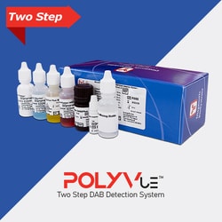
Diagnostic Biosystems POLYVUE PLUS MOUSE/RABBIT HRP Mouse/Rabbit PolyVue Plus™ HRP/DAB Detection System
The Mouse/Rabbit PolyVue Plus™ HRP/DAB Detection System is a non-biotin, two-step detection system suitable for labeling antigens in formalin-fixed paraffin-embedded tissues and cryostat sections. The PolyVue Plus™ Detection System may also be used with blood smears, cytosmears, and cell preparations. This system has been developed by directly labeling immunoglobulins with enzymes using a proprietary tandem hyperlabelling technology. This ensures consistent and reproducible immunostaining for all types of nuclear, cytoplasmic and membrane antigens in different types of tissues with significantly lower background than detection systems using biotin and avidin conjugates.

Diagnostic Biosystems ANTI-ISOCITRATE DEHYDROGENENAS Anti-Isocitrate Dehydrogenenase 1 (IDH1)-R132S
Isocitrate dehydrogenase 1 (IDH1) is a 46 kDa NADP-dependent enzyme, which catalyzes the decarboxylation of isocitrate into α-ketoglutarate. IDH1 may also play a role in the prevention of oxidative damage, and the shuttling of proteins to peroxisomes. It is widely reported that mutations in IDH1 result in multiple forms of gliomas. The Arginine to Serine substitution at amino acid 132 is seen in gliomas, abolishes magnesium binding and alters enzyme activity so that isocitrate is no longer converted to alphaketoglutarate but instead alpha-ketoglutarate is converted to R(-)-2- hydroxyglutarate.
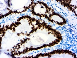
Diagnostic Biosystems SATB2 SATB2 (EP 281)
Special AT-rich sequence-binding protein 2 (SATB2) is a recently described marker that functions as a nuclear matrix-associated transcription factor. It has been reported that SATB2, in combination with CK20, could identify almost all colorectal carcinomas, including poorly differentiated colorectal carcinomas. Upper gastrointestinal (GI) carcinomas and pancreatic ductal carcinomas are usually negative for SATB2, and ovarian carcinomas, lung adenocarcinomas, and adenocarcinomas from other origin are rarely positive for SATB2. Therefore, SATB2 is a good marker for identifying a carcinoma of colorectal origin when working on a tumor of unknown primary. It can help differentiate colorectal metastasis (SATB2+) from primary pulmonary adenocarcinoma of mucinous or enteric type (SATB2- but often CDX2+)\n

Diagnostic Biosystems TISSUE BOND Tissue Bond™ Reagent
TissueBond reacts with silica glass to change its surface chemistry so that it may bond either ionically or covalently with aldehyde and ketone groups in tissue sections.\n\nProper tissue adhesion to glass slides is a critical step in immunohistochemistry. Good immunostaining may require harsh pretreatment of tissues for antigen retrieval (e.g. proteolytic enzyme digestion, boiling in high pH buffer, treatment with detergents or saponin). Improper adhesion of tissue sections can lead to poor quality staining or even loss of tissue sections. White glue, albumen, chrome-gelatin, and poly-L-lysine increase background staining. There is a need for a tissue adhesive that can irreversibly bind tissue sections to glass without creating background staining. Glass treated with TissueBond can be used for both paraffin and frozen tissue sections.
Diagnostic Biosystems DAB GONE KIT Montage DAB Gone™ Kit
Montage DAB GoneTM removes diaminobenzidine (DAB) stains and precipitate from surfaces. Contamination with DAB residue can be a problem that may affect staining quality or automated immunohistochemistry stainers. Bleach will only reduce the color intensity of DAB stains, whereas,Montage DAB GoneTM removes the residual DAB stain that may clog instrument probes and tubing. This kit can also be used to clean glassware and other labware used in the lab that has been exposed to DAB and has retained DAB stains even after normal cleaning. It chemically reacts with DAB to inactivate the carcinogenic properties.\n\nThis kit has been designed to clean DAB from Diagnostic BioSystems MontageTM 360. This kit can also be used with instruments such as: BioCare NemesisTM, intelliPATHTM, Dako Autostainer, Autostainer Plus, Autostainer Plus Link, Lab Vision AutostainerTM 360, 480, and 720 models.

Diagnostic Biosystems S-100 S100 (4C4.9)
S100 belongs to the family of calcium binding proteins and S100A and S100B are two members of the S100 family. S100A is composed of an alpha and beta chain whereas S100B is composed of two beta chains. This antibody stains with S-100 protein. S-100 protein has been found in normal melanocytes, Langerhans cells, histiocytes, chondrocytes, lipocytes, skeletal and cardiac muscle, Schwann cells, epithelial and myoepithelial cells of the breast,salivary and sweat glands, as well as glial cells. A large number of welldifferentiated tumors of salivary gland, adipose and cartilaginous tissue, and Schwann cell-derived tumors express S-100 protein. Almost all malignant melanomas and cases of histiocytosis X are positive for S-100 protein. S100 is of great value in the identification of several neoplasms, particularly melanomas and their metastases.

Diagnostic Biosystems CYTOKERATIN 19 Cytokeratin 19 (A53‐B/A2.26 )
This antibody is specific to a 40 kDa protein, cytokeratin 19. Its epitope maps between amino acids 312‐335. CK19 is expressed in sweat gland, mammary gland ductal and secretory cells, bile ducts, gastrointestinal tract,\nbladder urothelium, oral epithelia, esophagus, and ectocervical epithelium. Anti-CK19 reacts with a wide variety of epithelial malignancies including adenocarcinomas of the colon, stomach, pancreas, biliary tract, liver, and\nbreast. Perhaps the most useful application is the identification of thyroid carcinoma of the papillary type, although 50%-60% of follicular carcinomas are also labeled. Anti-CK19 is a useful marker for detection of tumor cells in lymph nodes, peripheral blood, bone marrow and breast cancer.